
The human skeleton contains 206 bones, all held in place by connective tissues such as ligaments and tendons. The skeleton gives structure to the body and protects the internal organs. Bones determine our size and shape. The skeleton also works as a factory, making various blood cells in the marrow of the bones. Bones also store minerals such as calcium and phosphorus for use by the body.
The structure of bones changes as children grow. The skeleton of the fetus in the womb is made up mostly of cartilage. As pregnancy continues, bone develops, but even when the child is born, there are still areas that are a combination of bone and cartilage. At the end of long bones, such as those in the arms and legs, there are growth plates. Growth plates are where new bone growth occurs and have a high level of activity until the child stops growing, usually around 13 to 15 years for girls and 15 to 17 years for boys.
Damage to the bones
Survivors of childhood cancer can develop a number of complications that involve the skeleton. These can be caused by surgery, radiation, and/or chemotherapy.
Amputation
If a malignant tumor extends to vital structures such as major nerves and blood vessels, then amputation (surgical removal of a limb) may be necessary. The missing limb will be replaced by an artificial body part, known as a prosthesis. Survivors who had only the lower parts of leg extremities (below the knee) amputated usually function well after rehabilitation, though functional and physical activities could be impaired by poorly fitted prostheses or pain. Upper or lower limb amputees may experience psychological problems and impaired quality of life that require professional support. Generally, amputees can ski, run, hike, and perform other physical skills very well.
One major decision I made with my parents was to choose a below-the-knee amputation instead of a second bone-salvage surgery. When first diagnosed, my treatment plan laid out by my oncologist and surgeon consisted of eight rounds of chemo, a bone-salvage surgery to remove my tibia (where the tumor was) and replace it with a donor bone, and then 10 more rounds of chemo following the surgery. A year after completing that treatment, the donor bone broke. My surgeon insisted that I undergo another bone-salvage surgery. This would have led to another 24-hour surgery, taking muscles and bone from other parts of my body to try and recover some semblance of function in my left leg, and this would still result in severe physical impairment for the rest of my life.
As an 11-year-old girl, I started doing independent research into other options. I discovered that amputation, while traumatic, would result in far better quality of life and allow me to go back to doing the things I loved, like dance and being active like a normal kid. My family and I decided to choose amputation below the knee, instead of the second bone-salvage surgery, and convinced my care team, even though it was not the original plan they hoped for.
Making these decisions and advocating for myself was never easy, but in the end resulted in the best quality of life for me. Fortunately, my support system and especially my parents allowed me to be a part of those decisions and gave me the confidence to continue to vocalize my needs and opinions when it comes to my continued long-term care.
These survivors may experience pain in the missing limb even though the limb is no longer present (known as phantom pain) and develop problems with calluses or pain in the stump. Those who had entire limbs removed or other portions of the skeleton removed (such as the pelvis) sometimes have ongoing problems with function and pain. Survivors requiring a hemipelvectomy (removal of all or part of the hemipelvis and the entire lower extremity) may have more functional problems.
The Amputee Coalition of America (ACA) provides extensive information about organizations and resources for amputees. https://www.amputee-coalition.org
Limb-salvage procedures
Growing numbers of survivors have limb-salvage procedures as an alternative to amputation. These procedures help save the limb by removing the part of the bone that is involved with the tumor. The bone is replaced with either a bone graft or a metal prosthesis. Another limb salvage procedure, known as rotationplasty, spares part of the lower leg so that the ankle can be rotated and used as a knee; below it, a prosthetic lower leg is surgically attached. In many cases, these surgeries are very successful, and the survivor’s limb works very well. Other survivors require multiple surgeries and cope with pain, infection, and functional and physical activity limitations.
Limb radiation
Radiation to a limb will impact growth if a child has not completed puberty. If the growth plate is included in the field of radiation, the untreated limb will continue to grow, creating a length discrepancy in the child’s limbs. This can be particularly problematic if the affected limb is a leg. The growth plate of the untreated leg may need to be surgically altered to stop growth to help keep the legs similar lengths.
In some cases, limb-lengthening procedures can reduce or correct the discrepancy in length between arms or legs while stretching the surrounding muscles and other soft tissues. This is a more involved process for the child and their family and can take six to nine months. The bone to be lengthened is cut and a lengthening device is surgically inserted between the cuts ends of the bone. In the next step, about a week later, the two ends of bone are slowly separated using the lengthening device. This is done by the family who have been trained by the surgeon how to use the device. This period, which usually takes a couple of months, is closely watched by the clinical team. After the limb has reached the desired length, the body takes over and new bone begins to grow and fill the gap.
Survivors who had radiation to a limb may be at risk for fractures without trauma, and fracture may occur after minor trauma that would otherwise not cause a fracture in an untreated bone. Fractures occur most often in children who received more than 4000 cGy to the bone. However, each survivor is different, so long-term follow-up is important.
Spine radiation
Some children with cancer get their entire spine irradiated. Others have a portion of the spine treated. Scoliosis, a sideways curvature of the spine (backbone), can be caused by receiving radiation to only one side of the spine that runs down the midline of the body. When the untreated side of the body continues to grow, the other side does not and the spine curves in the direction of the growing side (see Figure 18-2). Thankfully, children treated for cancer after 1990 will have a shorter trunk, but better spinal alignment. The damage is more pronounced for children treated when younger than age 6 years or during the growth spurt of puberty. A survivor whose whole spine was radiated also may develop scoliosis; this is thought to occur because damage to the muscles and soft tissue on the irradiated side of the body pull the spine out of alignment.
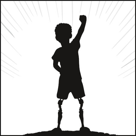
FIGURE 18-1. Survivors can continue to engage in physical activity and recreation, even run, hike, and ski. (©Alex’s Lemonade Stand Foundation, 2025)
Other factors that may increase the risk of developing scoliosis are changes to the spine from tumor, osteoporosis, and the surgical fusion of two parts of the spine to one another to treat pain.
A rare side effect of radiation to portions of the spine is kyphosis, where the upper spine curves outward, giving a hunchback appearance. This can occur with scoliosis after radiation to portions of the spine. Severe kyphosis and/or scoliosis can affect the functioning of other organs, such as the lungs, whose capacity can be reduced by the intrusion of the curved spine into the lung cavity.
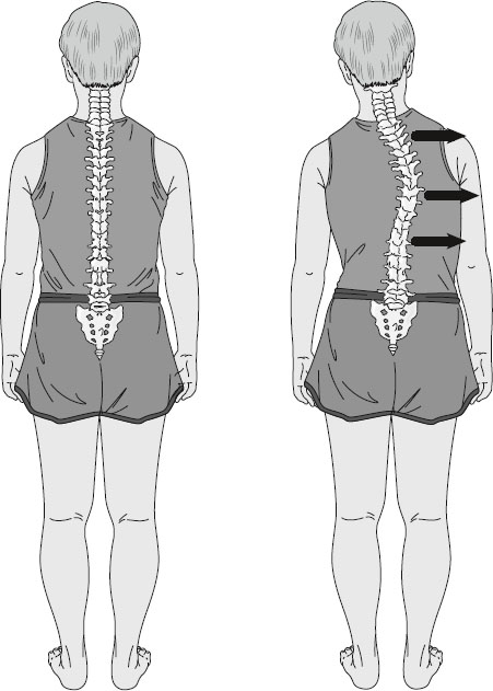
FIGURE 18-2. Scoliosis is curvature of the spine that occurs when one side of the body is irradiated and bones/muscles stop growing on one side of the body while the other side continues to grow causing the spine to shift and curve. (©Alex’s Lemonade Stand Foundation, 2025)
If radiation to the whole spine is needed, current protocols attempt to spare adjacent tissues to decrease the risk of scoliosis and kyphosis. Better staging of solid tumors has also decreased the number of children and adolescents who require radiation. Radiation to the whole spine can stop or slow the growth of the spine. A short trunk (measured from the top of the head to the rump) occurs most often in brain tumor survivors whose entire spines were radiated with more than 3500 cGy. Total body radiation given prior to stem cell transplantation (i.e., bone marrow, stem cell, or cord blood) can also affect the growth of the spine, as well as growth of other bones exposed to radiation.
Osteochondromas are outgrowths of the bone that are sometimes seen in children who were treated with radiation. These bony projections often form where bones meet one another and can also form on the bones of your spine. The outgrowths are also known as exostoses (singular form: exostosis) and are commonly referred to as bone spurs. Young children who receive total body irradiation (TBI) can develop osteochondromas as they begin puberty. An x-ray is needed to confirm the diagnosis. These growths often do not require intervention. However, in a small number of cases, removal is necessary due to location and discomfort.
Slipped capital femoral epiphysis (SCFE) is a disorder sometimes seen in children whose hip was irradiated. Epiphysis refers to the area at the top or head (capital) of the long bone of the thigh (femur) where it attaches to the pelvis. It has a wide and rounded shape and fits inside an area of the hip where the pelvic bones form a cup-like space. The epiphysis is separated from the rest of the thigh bone below it by a growth plate. When the growth plate is damaged and weakened by radiation, the head of the femur stays in the cup of the hip joint, but the rest of the thigh bone can “slip” relative to the femoral head. SCFE is often seen at the beginning of the pubertal growth spurt. Survivors who received radiation and who have endocrine dysfunction are at higher risk (Hobbie et al., 2022).
Osteoporosis and osteopenia
Some childhood cancer survivors develop osteoporosis, a disease that occurs when the creation of new bone doesn’t keep up with the loss of the old bone. When this happens, it causes the bone to lose density and to thin which make it weak, brittle, and more likely to break. This late effect (osteoporosis) is not well understood. It is known that survivors who took high doses of steroids (e.g., prednisone, dexamethasone), received high-dose methotrexate, had cranial radiation, had high-dose radiation to bones, or have low growth hormone seem to be most at risk. Young women who have an early menopause and men with low testosterone production also have a higher risk of developing osteoporosis.
Osteopenia is similar to osteoporosis but not as serious. Like survivors with osteoporosis, those with osteopenia have a lower-than-normal bone density and experience a thinning and weakening of their bones. While the bones are not as strong as they should be, the loss of bone is not as great as it is in osteoporosis. Osteopenia can develop into osteoporosis.
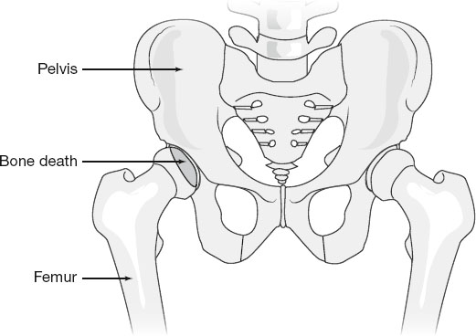
FIGURE 18-3. Osteonecrosis, or bone death, in the hip. (©Alex’s Lemonade Stand Foundation, 2025)
Osteonecrosis
Osteonecrosis is the death of bone tissue due to a lack of blood supply which can lead to tiny breaks in the bone and cause the bone to collapse (see Figure 18.3). Other names for this condition are avascular necrosis (AVN) and ischemic necrosis. It is usually caused by radiation to bones and/or use of high-dose steroids (e.g., prednisone or dexamethasone).
Osteonecrosis is generally seen early rather than late—often within the first year after therapy. Adolescent girls are especially susceptible. Magnetic resonance imaging (MRI) is the best tool for diagnosis. As bone deterioration progresses, the bone may become weak and may eventually collapse. The course of the disease is variable. Some survivors have osteonecrosis for years with only minor problems with pain or movement, while others require surgery soon after diagnosis. Osteonecrosis can be very painful and sometimes leads to osteoarthritis (see below). The website for the Avascular Necrosis Support Group is located at www.facebook.com/groups/AVNInfo/.
Osteoarthritis
Osteoarthritis is a degenerative disease of the joints and occurs when the cartilage that protects and cushions the ends of bones wears down over time. It is characterized by pain with activity that subsides when resting. Survivors who had radiation to joints are at risk. People who have late effects that increase stress on the joints, such as osteonecrosis, may also develop osteoarthritis. Helpful information about arthritis can be found at the Arthritis Foundation’s website—www.arthritis.org
Signs and symptoms of damage to the bones
Leg length discrepancy:
Difference in length and muscle mass between two limbs
Limping
Lower back pain
Hip pain
Scoliosis
Fractures:
Pain
Deformity
Swelling
Bruising
Loss of function
Scoliosis and kyphosis:
Curved spine
Uneven shoulder height
Back or hip pain
Limping
Spinal shortening
Osteonecrosis, osteoarthritis, and SCFE:
Pain in affected joint
Limping
Impaired function
Osteochondromas:
Hard lump on any boney surface
Possible pain (depending on location)
Osteoporosis and osteopenia usually have no signs or symptoms, and bone density changes might not show up on regular x-rays unless the affected bone gets broken or infected. Advanced osteoporosis can cause the bones in your spine to compress, causing shorter height. It can also cause kyphosis (a hump on the upper part of the back), discussed earlier.
Follow-up screening and detection of bone damage
Amputation or limb salvage
Survivors with amputations need an annual examination of the stump that includes a discussion about whether a prosthesis could be useful. Range of motion and function are evaluated. Survivors who had a limb salvage procedure should have both treated and untreated limbs measured every year (without clothes on to get accurate measurements), usually by an orthopedic surgeon. An orthopedic surgeon is a doctor who specializes in diagnosing and treating disorders involving the bones, joints, ligaments, tendons, and muscles, particularly performing surgery. A baseline and then annual x-rays are needed until growth is complete to assess the growth plate. Survivors should also have a discussion with their healthcare provider if they have any back pain, limb pain, limping, or changes in muscle mass.
As a below-the-knee amputee, a critical part to my physical mobility is my prosthesis. Unfortunately, high quality and customized prosthetics are poorly covered by insurance policies in the United States. There really is not a good option that provides amputees with quality prosthetics without paying enormous costs out of pocket for them. I have been very fortunate to have help from my family with covering those out-of-pocket costs to date, but there will come a time where I will be paying 5 figure costs for my prosthesis unless policies change dramatically. This is a huge ongoing issue, and a key focus of many advocates for amputees nationwide.
Scoliosis and kyphosis
A scoliosis check needs to be done every year if you are at risk. Your back will be examined while you bend over with your fingers touching your toes and your knees straight. Kyphosis is often seen on observation, but should be confirmed using X-rays.
If your child had radiation to the spine, several tests should be done to check for spinal abnormalities. Your child should have height checked while both standing and sitting, and the results should be plotted on a chart. Your child’s healthcare provider should examine your child’s spine every 3 months during puberty until growth is complete, and every year thereafter. A spine x-ray should be obtained for a baseline before puberty, then as needed.
Osteopenia, osteoporosis, and fractures
If you had more than 4000 cGy radiation to any bones, they are at risk for developing osteopenia and osteoporosis which can lead to fractures. Go to your healthcare provider if you have pain, swelling, or bruising in the areas that were irradiated. You may also be at risk if:
You went into an early menopause (your periods stopped early).
You are a stem cell transplant survivor who took high doses of steroids.
You have any ovarian or testicular dysfunction.
You have growth hormone deficiency.
You have a family history of osteoporosis.
Discuss your risk with your healthcare provider to see whether you need bone density studies. X-rays or computed tomography (CT) scans may be done to assess the amount of damage.
A painful joint should be evaluated by your healthcare provider. You may need an x-ray or CT scan to check for osteoarthritis and osteonecrosis.
Medical management of bone damage
Amputation or limb salvage
Medical management of amputations includes a visual inspection and checks for range of motion and muscle contractures. You should have your prosthesis checked (if you use one) and have a discussion with your healthcare provider about any advancements in technology.
If you had limb salvage surgery or radiation and one limb is now longer than the other, discuss the treatment plan with your healthcare provider. Small differences in length of less than 2 centimeters (cm) usually don’t require any treatment. Differences of 2-6 cm are treated with a shoe lift or surgery to stop the growth of the other limb. If the discrepancy is greater than 6 cm, other surgical steps may be necessary. These include shortening the untreated limb or lengthening the treated leg to restore a comfortable gait.
If you have an endoprosthesis, which is an artificial body part that is placed entirely inside your body (such as an artificial hip), you need to take antibiotics prior to dental work to prevent possible infection that could spread to the prosthesis.
Scoliosis and kyphosis
If you have any scoliosis or kyphosis, you should be referred to an orthopedic specialist with experience treating survivors of childhood cancer. If the curvature is noted during a period of rapid growth, such as puberty, do not delay seeing the specialist, because the curvature can increase rapidly during a growth spurt. Long-term survivors who have scoliosis or kyphosis often have back pain.
The following may help make the pain more manageable:
Physical therapy
Using a brace
Moderate exercise
Pain medication
If the curve in the back progresses (worsens) beyond 30 degrees (or curves 20 degrees with rapid progression), wearing a brace may be necessary. Curves greater than 40 degrees may require surgery. There are no standards for treatment of scoliosis after tumor surgery or radiation; the decision to use a brace or operate is performed based on the survivor’s specific needs. Kyphosis treatment includes the use of any pain relievers as needed and exercises to strengthen the musculature around the spine. Possibly bracing may be used, but may not be an option for survivors. Surgical options are reserved for survivors with severe functional impairments.
Osteoporosis or osteopenia
If you have osteopenia or osteoporosis, your yearly examination should include education on the importance of:
Weight-bearing exercise such as walking or running.
A diet rich in calcium. This includes dairy products, shellfish, leafy green vegetables, and tofu. Your healthcare provider might also recommend taking supplemental calcium with added vitamin D.
Adequate vitamin D. Some dairy products are fortified with vitamin D and the body makes some on its own when exposed to sunlight, but many survivors require supplementation. A nutritionist consultation may be helpful especially in the case of food allergies or to determine the best nutritional supplements.
Osteonecrosis
Treatment for osteonecrosis may include:
Activity modifications
Range of motion exercises
Electrical stimulation
Surgery to remove the inner layer of bone (core decompression) that involves drilling into the area of dead bone near the joint to reduce pressure and increase blood flow; sometimes a healthy piece of bone is inserted when the inner layer is removed. This procedure may slow or stop the osteonecrosis.
Surgery to cut and realign a bone near a damaged joint (osteotomy) to reduce the weight bearing of the bone with osteonecrosis
Pain and/or anti-inflammatory medication
Fractures
If you have a fracture in irradiated bone, most likely the fracture will occur where the bone was biopsied or at the site of tumor in the bone. These types of fractures may need surgery to insert a device to keep the bone aligned. Because the surrounding tissues were also irradiated, this surgery will be challenging. Make sure the surgery is done by an orthopedic surgeon with experience operating on irradiated bone and tissues.
Osteochondromas
Osteochondromas are generally noted first by the survivor or a family member. Once the diagnosis is confirmed by x-ray, management will be determined by the symptoms. If there is pain or if the osteochondroma is affecting other bone growth or joint function, a referral to an orthopedist is recommended.
Table of Contents
All Guides- Acknowledgements
- Contributors
- Foreword
- Preface
- 1. Survivorship
- 2. Emotions
- 3. Relationships
- 4. Navigating The System
- 5. Staying Healthy
- 6. Genetic Testing And Childhood Cancer
- 7. Diseases
- 8. Fatigue
- 9. Brain And Nerves
- 10. Hormone-Producing Glands
- 11. Eyes And Ears
- 12. Head And Neck
- 13. Heart And Blood Vessels
- 14. Lungs
- 15. Kidneys, Bladder, And Genitals
- 16. Liver, Stomach, And Intestines
- 17. Immune System
- 18. Muscles And Bones
- 19. Skin, Breasts, And Hair
- 20. Subsequent Malignancies
- About The Editors