
Childhood Cancer
The eye
Each structure within the eye has a specific task to help transmit information from the outside world through the optic nerve to the brain (see Figure 5-1).
The cornea is the curved outer portion of the eyeball that transmits light to the retina. Behind the cornea is the iris, which is the colored portion of the eye. The iris controls the amount of light entering the eye by making the opening at the center, called the pupil, either larger or smaller.
As light rays pass through the curved surface of the cornea, they are bent and then passed through the pupil. Inside the eye, sitting behind the pupil, is a disc-shaped structure called the lens. The lens is clear in a healthy eye, and it has two curved surfaces that refract light two more times on its journey to the back of the eye. As light rays travel from the front to the back of the eye, they pass through a colorless, jelly-like material, called the vitreous, which fills the eyeball behind the lens.
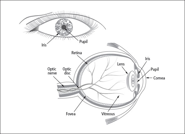
Figure 5-1: The anatomy of the eye
The inside lining of the eyeball consists of a structure called the retina. The retina converts light energy into electrical impulses that are transmitted to the brain. The fovea is the area of the retina that is the center of vision, and the optic disc is the area of the retina where the optic nerve enters the eye. Retinoblastoma tumors develop in the retina of the eye because of a genetic mutation, either inherited or acquired. This mutation causes abnormal growth of immature retinal cells called retinoblasts.
Thirty years ago, when I was 18 months old, I was diagnosed with retinoblastoma. I had one tumor in just one eye. The eye was surgically removed, and I had no chemotherapy or radiation. I have no late effects other than wearing a prosthesis and having vision in only one eye.
Table of Contents
All Guides- Introduction
- 1. Diagnosis
- 2. Bone Sarcomas
- 3. Liver Cancers
- 4. Neuroblastoma
- 5. Retinoblastoma
- 6. Soft Tissue Sarcomas
- 7. Kidney Tumors
- 8. Telling Your Child and Others
- 9. Choosing a Treatment
- 10. Coping with Procedures
- 11. Forming a Partnership with the Medical Team
- 12. Hospitalization
- 13. Venous Catheters
- 14. Surgery
- 15. Chemotherapy
- 16. Common Side Effects of Treatment
- 17. Radiation Therapy
- 18. Stem Cell Transplantation
- 19. Siblings
- 20. Family and Friends
- 21. Communication and Behavior
- 22. School
- 23. Sources of Support
- 24. Nutrition
- 25. Medical and Financial Record-keeping
- 26. End of Treatment and Beyond
- 27. Recurrence
- 28. Death and Bereavement
- Appendix A. Blood Tests and What They Mean
- Appendix B. Resource Organizations
- Appendix C. Books, Websites, and Support Groups