
The eye is the organ of sight. This nearly spherical body has numerous layers. At the front is the transparent cornea; through the cornea, the colored iris is visible. The space between the cornea and the iris is the anterior chamber, which is filled with a clear fluid called aqueous humor. In the center of the iris is a space called the pupil that contracts or expands to control the amount of light entering the eye. Directly behind the pupil is the lens of the eye. The cavity behind the lens is filled with a gel-like substance called the vitreous humor. The thin layer that lines this cavity is the retina which detects the light signals that enter the eye. The optic nerve transmits light information from the retina to the brain. Figure 11-1 shows the anatomy of the eye.
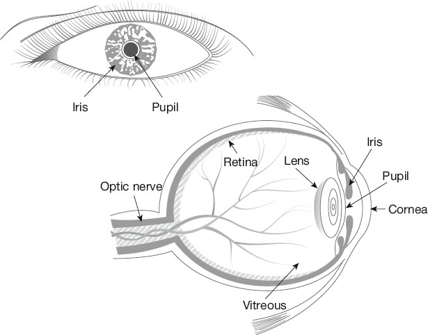
FIGURE 11-1. Anatomy of the eye. (©Alexʼs Lemonade Stand Foundation, 2025)
Both of our girls had 6 months of VEC chemotherapy and PRN laser/cryotherapy. They have both had chemotherapy injections into their eyes. Our oldest had 3 rounds of IAC at Jefferson. All of this to shrink and kill the tumors on their retinas and prevent them from spreading to the brain. One of our girls will only have peripheral vision in her left eye with close to normal vision in her right. The other will likely have normal vision in both eyes.
Eye damage
The eye can be damaged by radiation, treatment with steroids, or surgery. Vision can also be harmed if the optic nerve is damaged anywhere between the eye and the brain which can happen from radiation, a tumor pressing on the optic nerve, or a tumor in the brain causing high fluid pressure around the brain and optic nerves. In addition, tumors in the brainstem (where the brain meets the spinal cord) can cause problems with eye movement or double vision.
When a cancer is in the eye, as in retinoblastoma, the affected eye might have to be surgically removed (called enucleation); this occurs less often now than in the past because of newer radiation techniques and chemotherapy protocols. Small and medium-sized retinoblastomas are often treated with some combination of chemotherapy, cryotherapy (freezing treatment), and photocoagulation (laser treatment). Large or advanced tumors may require enucleation. See more information about retinoblastoma on website of American Academy of Ophthalmology: https://www.aao.org/eye-health/diseases/what-is-retinoblastoma
The eye is exposed to low-dose radiation during total body irradiation (TBI) prior to some types of stem cell transplants. High-dose radiation is used to treat retinoblastoma, rhabdomyosarcoma, and other tumors around the eye. The eye may also be exposed to radiation given to treat brain tumors or leukemia.
Common problems of the eye after treatment
Hypoplasia (slowed growth of the bones around the eye)
Dry eye
Shrinkage of the lacrimal duct, which drains tears from the eye
Loss of vision
Cataracts
A common late effect is slowed growth (hypoplasia) of the orbits (eye sockets), which are the bony structures in the skull that protect the eyes. Each orbit is made up of seven bones, all of which can be damaged by radiation. This late effect is most noticeable in survivors who were treated at a very young age with more than 4000 cGy in the areas of the growing bones. Survivors with severe hypoplasia sometimes benefit from reconstructive surgery after the bones in the face are fully grown.
Cataracts (when the lens of the eye becomes opaque) are a frequent late effect of radiation treatment. Cataracts can cause vision loss, but they can often be treated with surgery. Higher doses of radiation cause more damage and shorten the time before cataracts appear. Cataracts appear earlier and become more severe if the child was treated at a young age. In addition, single, larger doses of radiation cause cataract formation more often than the same total dose given in several smaller fractions. Long-term steroid use (including prednisone and dexamethasone) or busulfan chemotherapy can also cause cataracts. See more information on cataracts on the American Academy of Ophthalmology website: https://www.aao.org Search: what are cataracts.
Graft-versus-host disease and/or radiation can cause mild to severely dry eyes. Symptoms include irritation, burning, pain, blurred vision, sensitivity to light, and the feeling that a foreign body is in the eye. Dry eyes can be eased by using artificial tears.
Loss of vision can involve central vision (the detailed vision used for reading) or the peripheral visual fields (side vision). Loss of peripheral vision can result from damage to the retina or optic nerve or damage to the visual areas of the brain from brain surgery.
Less common late effects to the eye or eye socket from radiation can include:
Permanent loss of the eyelashes
Hemorrhaging of small damaged blood vessels in the retina (telangiectasias) typically occurring 3 to 5 years after treatment
Adhesions and scarring that limit the ability of the lids to fully close, making the eyes drier
Inflammation of the cornea (called keratitis), with sensitivity to light and pain at the surface of the eye
Ulcers, holes, or scarring of the cornea after treatment
Glaucoma (increased pressure within the eye) which can also be caused by steroid use
Secondary cancers
Surgery, stroke, and some medications can also cause problems with the eyes and vision. If there is damage to the part of the brain that controls the muscles that move the eyes, this can result in an eye that does not move normally, and the two eyes not being lined up with one another. If this happens at an early age, the brain may start to ignore one eye, and the eye can lose vision as a result (called amblyopia). Amblyopia may need to be treated with patching therapy (patching the good eye to improve vision in the eye that is not being used). Surgery may help improve the alignment of the eyes, but surgery cannot always restore a full range of movement.
Signs and symptoms of eye damage
When children are old enough to talk, and certainly by school age, visual disturbances are usually detected and treated. However, if changes are gradual, a child may get used to them, and identification of visual late effects can be delayed.
The following are signs and symptoms of late effects to the eye:
Blurry vision
Double vision
Blind spots or a decrease in visual field (range of vision)
Increased sensitivity to light
Difficulty with night vision
Floaters across the field of vision that change or light up
Pain in the eye
Decreased tearing or excessive tearing/watering of eyes
Persistent dry, scratchy eyes or eyelids
Follow-up screening and detection for eye damage
You should be seen regularly by an ophthalmologist with experience treating survivors of childhood cancer if you:
Had total body radiation
Received high-dose radiation to one or both of the eyes or nearby structures
Received radiation for a brain tumor or leukemia that included an eye in the radiation field
Used steroids for a prolonged period
The ophthalmologist should perform a thorough evaluation that includes evaluation of vision, a test for glaucoma, checking for cataracts, and examination of the retina.
Survivors not at high risk for eye damage will usually have a brief eye examination as part of their yearly follow-up examination. This should include a visual examination of the eyes and a vision test using a Snellen letter chart, or a picture chart for children who cannot read letters. Even children as young as age 3 can identify simple pictures on charts specially designed for preschoolers. Any abnormalities should result in a referral to an ophthalmologist.
For additional information about vision tests you should have based on your specific treatment, you and your healthcare provider can refer to the Children’s Oncology Group’s survivorship guidelines at www.survivorshipguidelines.org.
Medical management of eye damage
If you have any visual changes, you should be followed by an ophthalmologist skilled in treating survivors of childhood cancer. If you have cataracts or glaucoma, you may need glasses, medications, and/or surgery. Survivors who had one or both eyes removed will need a series of well-fitted prostheses as they grow, and education about caring for the prostheses.
A survivor’s healthcare team should also provide referrals to resources to assist with adjustments related to limited vision. Special arrangements should be made with schools to provide preferential seating, eyewear protection, and other necessary accommodations.
Childhood survivorship clinics or programs can provide detailed information about educational regulations and opportunities for young people with visual impairments. For more information about education, see Chapter 4, Navigating the System.
Artificial tears and ointments are used to protect the cornea and preserve vision in survivors with chronic dry eyes. A severe problem with dry eyes can sometimes be fixed by closing the tear draining ducts with either plugs or surgery (called punctal ligation). It also helps to avoid anything that contributes to dry eyes such as wind, fans, smoke, low humidity, or air conditioning.
Survivors who develop corneal ulcerations as complications of dry eyes may need to be treated with antibiotic eye drops.
Radiation-induced cataracts are progressive. Exposing these cataracts to extra sunlight may speed the progression. Sunlight can also aggravate glare symptoms from cataracts. Wearing sunglasses with UV protection when outside may help children and teens with cataracts.
Table of Contents
All Guides- Acknowledgements
- Contributors
- Foreword
- Preface
- 1. Survivorship
- 2. Emotions
- 3. Relationships
- 4. Navigating The System
- 5. Staying Healthy
- 6. Genetic Testing And Childhood Cancer
- 7. Diseases
- 8. Fatigue
- 9. Brain And Nerves
- 10. Hormone-Producing Glands
- 11. Eyes And Ears
- 12. Head And Neck
- 13. Heart And Blood Vessels
- 14. Lungs
- 15. Kidneys, Bladder, And Genitals
- 16. Liver, Stomach, And Intestines
- 17. Immune System
- 18. Muscles And Bones
- 19. Skin, Breasts, And Hair
- 20. Subsequent Malignancies
- About The Editors